Dry Needling for Plantar Fasciitis: Thoughts for Treatment
Dry Needling for Plantar Fasciitis: Thoughts for Treatment
Plantar Fasciitis (PF) can be one of those annoying, chronic conditions to deal with, both as a patient and as a practitioner. First of all, the term PF has basically become nonsensical by such widespread misuse. Pretty much any foot pain is diagnosed as plantar fasciitis. We have all seen this. Therefore, and unfortunately, many plans of care following the diagnosis of PF include the foot, and only the foot.
What causes Plantar Fasciitis
Aside from some structural anomaly or specific injury in the foot, it is exceedingly rare that the cause of PF lies soley in the foot. The following anomalies can cause PF:
- pelvic
- calf
- knee
- hamstring
- spine
- pelvic floor
Yes, even the pelvic floor, so now you get the idea. A lot of things can cause PF. This is not to say that I do not treat the foot, because it needs to be treated, sometimes. I have personally only had to deal with plantar foot pain (that should be the name instead of PF) a couple of times, and it hurts!
However, in most cases, in the absence of specific foot pathology, the pain in the foot should be treated as a symptom, not the cause. If it is treated as the cause, and other parts of the body are not addressed, the vast majority of the time the foot pain will return. This is exactly why this is a chronic condition with so many people because the cause of the problem is never addressed. It may improve only if the foot is addressed, but if it returns, something else is likely causing the problem.
How Tibialis Posterior impacts PF
I have found the tibialis posterior to be one of the most problematic muscles when it comes to foot pain. It starts way up on the back of the proximal tibia, travels down the posterior tibia, and the tendon wraps around the medial malleolus and attaches to numerous bones in the foot. The tib posterior directly affects the arch of the foot more than any other muscle because of its size, strength, and anatomical orientation. It has a long lever arm and a short force arm after it wraps around the medial malleolus. This gives the muscle a significant mechanical advantage. Kinda like superpowers. I really like superpowers.
Dry Needling Treatment for PF
The following is from the University of Washington, Department of Radiology.
Origin: Posterior aspect of interosseous membrane, superior 2/3 of medial posterior surface of fibula, superior aspect of posterior surface of tibia, and from intermuscular septum between muscles of posterior compartment and deep transverse septum
Insertion: Splits into two slips after passing inferior to plantar calcaneonavicular ligament; superficial slip inserts on the tuberosity of the navicular bone and sometimes medial cuneiform; deeper slip divides again into slips inserting on plantar surfaces of metatarsals 2 – 4 and second cuneiform
Action: Principal invertor of foot; also adducts foot, plantar flexes ankle, and helps to supinate the foot
Innervation: Tibial nerve (L4, L5)
Arterial Supply: Muscular branches of sural, peroneal and posterior tibial arteries
This is a difficult muscle to treat directly without needles. Actually, it’s impossible. Have you ever thought about the fact that aside from internal exams, mouth, other stuff, it is impossible to directly treat any muscle in the body without breaking the skin? This is one of the many reasons why DN, properly performed, is incredibly powerful. Aside from the skin, which is certainly important, it is the only direct treatment of most tissues we have available to us.
Related: Click here to learn more about our Dry Needling Courses
It is important to consider the spatial orientation of the tibial nerve, posterior tibial artery, and vein in relation to the tib posterior. These neurovascular structures basically run down the center of the posterior aspect of the tib posterior. If the tib posterior shortens and, by default, thickens, the arch will be elevated and the neurovascular bundle will become compressed between the tib posterior and the soleus to some degree. When this happens, a number of cascading events follow: microvascular circulation in the tib posterior decreases and less blood is available, leading to hypoxia of the muscle and nerve, increased chemical concentrations ensue, pain is amplified, etc.
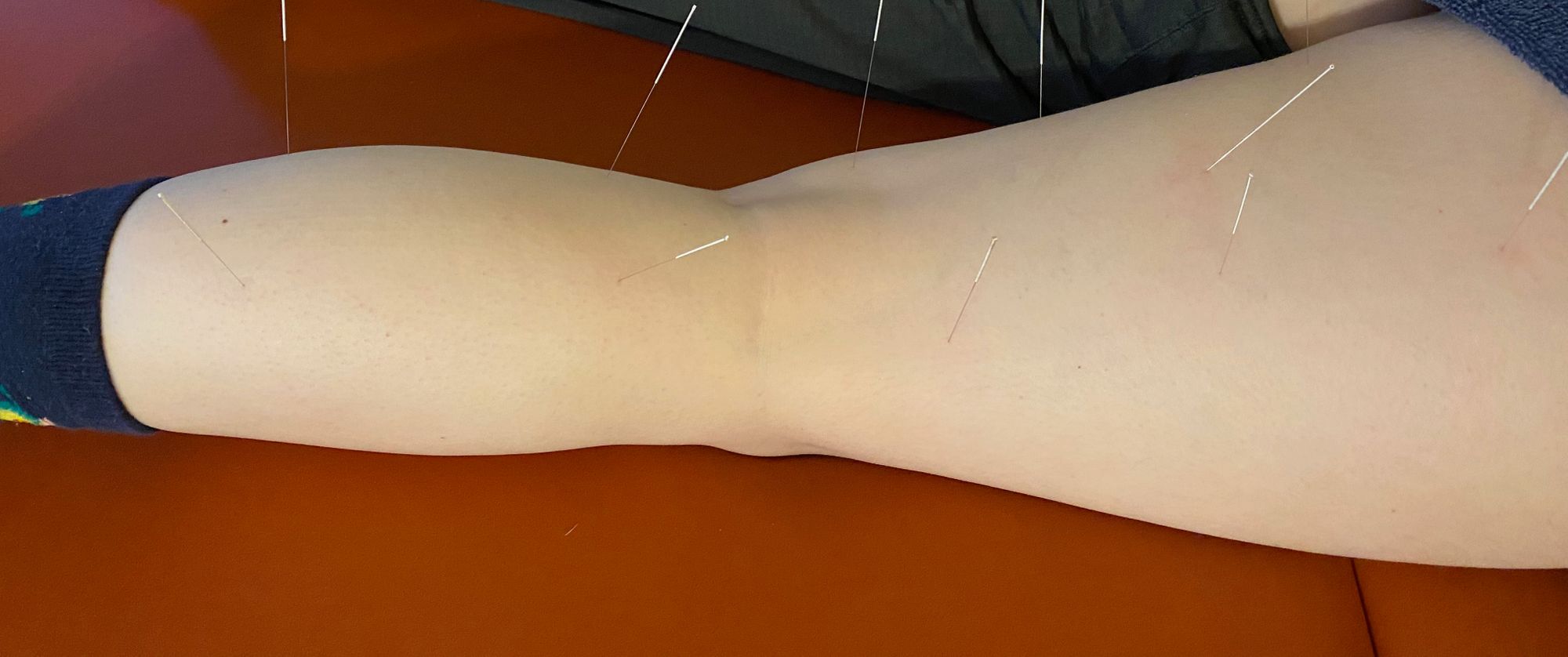
The way I typically access the tib posterior is at the medial aspect of the tibia somewhere in the distal half of the muscle. This is the easiest and most comfortable area to access this muscle when I needle myself and from clinical experience. It is also an area that tends to be pathologic, which is handy, like a pocket on a shirt. It is typically the first significant tissue resistance felt with the needle if it is placed properly, and the patient usually will let you know by wincing for a second. This may also cause an aching pain in the foot. Remember, it is difficult to hit nerves on purpose without imaging. This nervy sensation is typically from the muscle, or the muscle contracting or fasciculating and putting intermittent pressure on the tibial nerve, or referred pain. One of the reasons I do not like to “piston” needles, in general, is that not doing so allows you to know whether or not you hit a nerve. And if you do happen to hit a nerve, which is highly unlikely if you are paying attention at all, you only hit it once. Same goes for pleural tissue. The only time I piston a needle is when I get a twitch with an initial insertion. Then I get all the twitches out. Unless it is the first time I am treating someone, then I try to not get twitches, and if I do, I do not attempt to get more. It is good to allow the patient and their nervous system to slowly adjust to the experience of DN.
When you are inserting a needle towards an area where a significant nerve typically hangs out, if you think you have hit a nerve and you are not moving the needle in and out of the tissue, just leave the needle there for a few seconds. If the pain subsides at all, which it does 99.99% of the time, you know you have not hit the nerve. If you actually did hit the nerve, there is no harm leaving this tiny needle there for a second. If you do not leave the needle there to see what happens, you will never know whether or not you hit a nerve. Remember that the gold standard ANS test, microneurography, involves inserting a solid needle directly into a nerve, typically the common peroneal.
Remember, nerves need fresh blood just like everything else in our bodies. This decreased circulation, especially decreased venous return, which is the most affected, allows for elevated concentrations of certain substances, such as calcium, acetylcholine and carbon dioxide, along with depressed levels of other substances, such as acetylcholinesterase, ATP, and oxygen. When this happens, a negative feedback loop begins: more pain, tighter muscles, less blood flow, less oxygen, more chemicals, more pain, and round and round it goes. DN is, by far, the fastest way I know how to stop and reverse this process to achieve a more homeostatic condition.
As far as DN goes, another awesome reason to not begin in the foot, unless otherwise indicated, and no, foot pain is not necessarily an indication, is that it freaking hurts. Hey, would you like a needle stuck into your foot? Does that sound like a good question to ask a new patient or a patient hesitant to try DN? Ha, ha. Unless the patient is in a real hurry to get better, I never begin treatment by needling the foot. It is just not that comfortable, although it can be extremely effective. The most therapeutic way to treat PF or foot pain is to treat from the spine to the foot all at the same time and connect it with stim. I like 1-5 Hz, which is best at endogenous opioid release, beta-endorphin in particular. But needling the foot hurts, and you can fix a large percentage of foot pain without actually needling the foot. I’m not saying don’t treat the foot, just don’t treat it right off the bat with needles. Your patients will like you better, I promise.
Related: Why you need both DN and Manipulation
There are a lot of simple and effective foot and ankle manipulations that we teach in our extremity manipulation course that I consistently use with my patients. This is a good technique to implement in place of DN, at least off the bat. This is a good example of how helpful it is to use DN and manipulation in conjunction. I think those are the two most powerful tools we have at our disposal to make significant and lasting changes on various neuromusculoskeletal systems.
DISCLAIMER: The content on the blog for Intricate Art Spine & Body Solutions, LLC is for educational and informational purposes only, and is not intended as medical advice. The information contained in this blog should not be used to diagnose, treat or prevent any disease or health illness. Any reliance you place on such information is therefore strictly at your own risk. Please consult with your physician or other qualified healthcare professional before acting on any information presented here.
Stay Engaged With Intricate Art
Get the latest news, updates and offers from Intricate Art delivered to your inbox.
